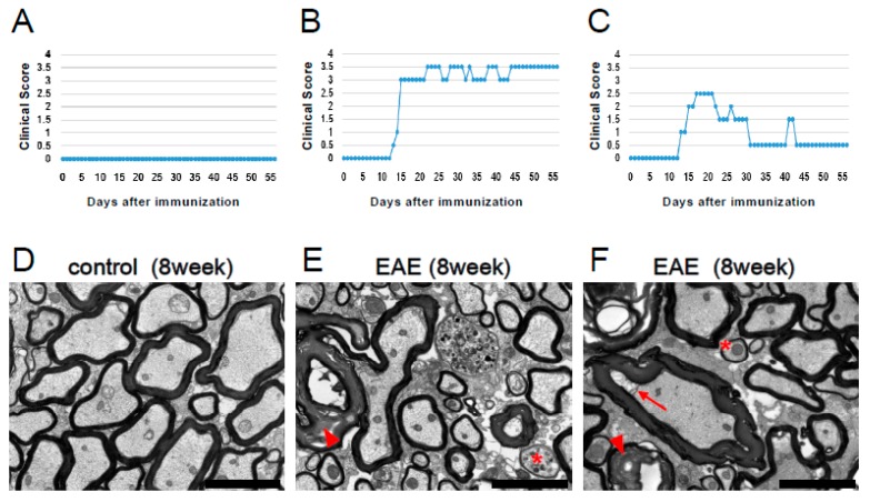
Figure 4.
Clinical scores (A–C) and EM findings (D–F) in control (A, D) and MOG-injected EAE mice (B, C, E, and F) that either retained high clinical scores (B, E) or showed an improvement in clinical scores (C, F). Axons and myelin were intact in the lumbar spinal cord of control mice (D). In contrast, both demyelination (excess formation of myelin [arrowheads in E and F], detachment of myelin from axon [arrow in F]) and remyelination (asterisks in E and F) were observed in lumbar spinal cords after EAE induction. Scale bars = 3 µm (D–F). Abbreviations: EM, electron microscopy; MOG, myelin oligodendrocyte glycoprotein; EAE, experimental autoimmune encephalomyelitis.

