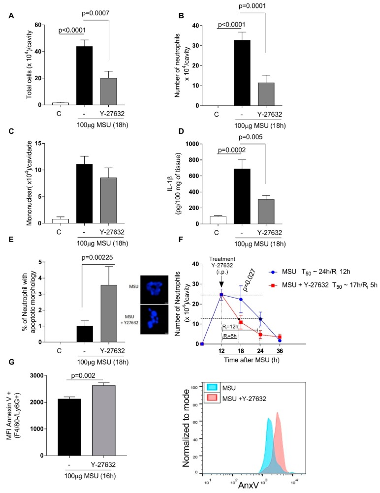
Figure 4.
Effects of Y-27632 treatment on Monosodium Urate Crystals (MSU) - induced gout. C57BL/6J mice were injected with MSU crystals (100 μg) into the tibiofemoral joint and 12 h later received an injection of Y-27632 (10 mg/kg i.p.). Knee were washed 6 h after treatment to cell count total number cells (A), number of neutrophils (B) and number of mononuclear cells (C). Levels of IL-1β (D) were measured by ELISA and expressed in pg/100mg of periarticular tissue. Cells with distinctive apoptotic morphology were determined 6 h after treatment (E). Resolution indices were quantified (F). Of note, Tmax = 12 h, the time point when polymorphonuclear (PMN) numbers reach maximum; T50 Y-27632 ~17 h, the time point when PMN numbers reduce to 50% of maximum; and Ri Y-27632 ~5 h; Ri, the time period when 50% PMNs are lost from the knee cavity. Apoptosis was biochemically 4 h after Y-27632 treatment and the median of fluorescence intensity (MFI) of annexin V was determined by flow cytometer (G). Data were collected with FACSCanto II flow cytometer and analyzed with FlowJo software. Data are shown as the mean ± SEM of five mice in each group (ANOVA test followed by Holm-Sidak’s multiple comparison).

