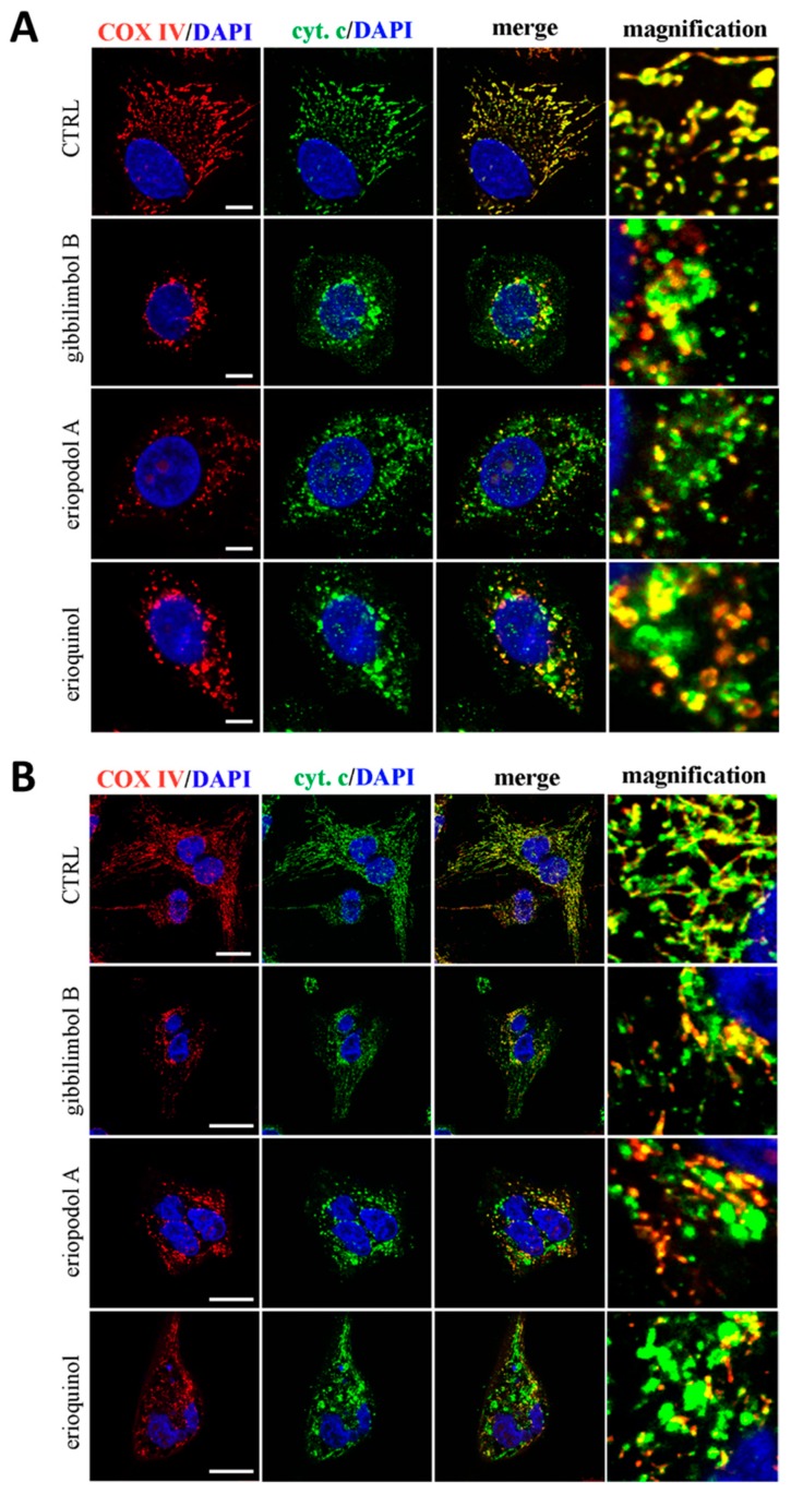
Figure 8.
Confocal microscopy for co-localisation of cytochrome c with mitochondria. (A) MCF7 and (B) U373 cells were treated for 3 h in the absence (CTRL, control) and in the presence of gibbilimbol B/eriopodol A (30 µg/mL) or erioquinol (10 µg/mL). Cells were then stained for cytochrome c (green) and the mitochondrial marker COX IV). 4’,6-diamidine-2’-phenylindole dihydrochloride (DAPI) (blue) was used for nuclei detection. The images are representative of three independent experiments. Scale bars: 10 µm (MCF7) and 25 µm (U373). Panels on the right represent enlarged image details.

