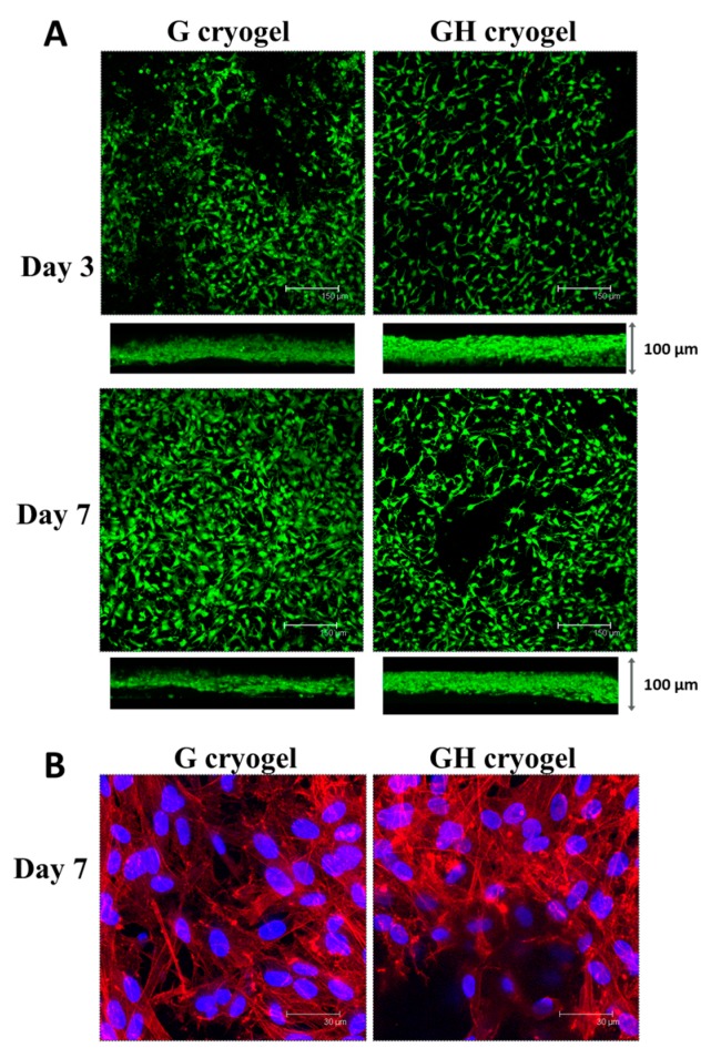
Figure 5.
Confocal microscopy observation of mesothelial cells cultured in G and GH by live/dead (A) (bar = 150 μm) and nucleus/cytoskeleton staining (B) (bar = 30 μm). The live cells were stained green and the dead cells were stained red in (A), while the cell nuclei were stained blue by Hoechst 33342 and the actin cytoskeleton was stained red by rhodamine-phalloidin in (B). Both the merged top-view image and cross-sectional-view image are included in (A).

