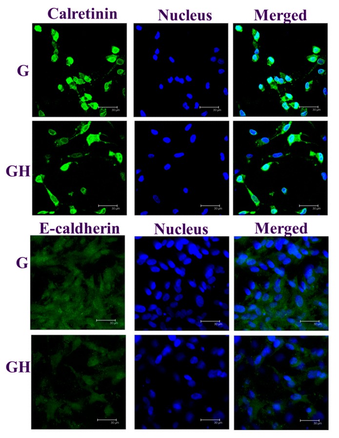
Figure 7.
The immunofluorescence (IF) staining of calretinin and E-cadherin of the mesothelial cells cultured in G and GH for seven days. The protein was stained green by a fluorescein isothiocyanate (FITC)-conjugated secondary antibody, while the nuclei were stained blue by Hoechst 33342. Bar = 30 μm.

