Figure 2.
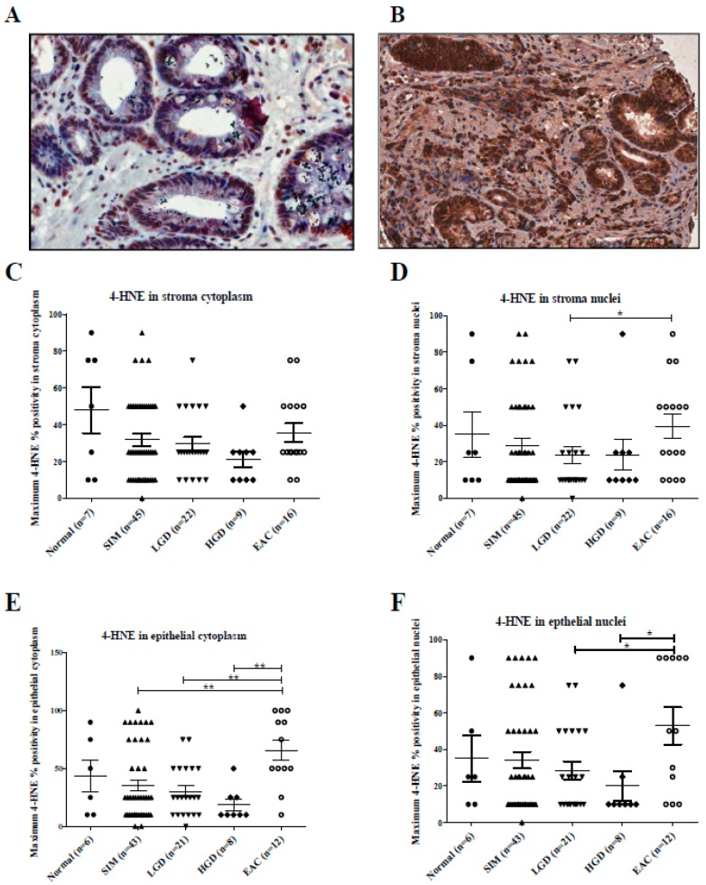
(A–F): 4-HNE stroma and epithelial staining in aged-matched histology groups across the Barrett’s esophagus disease spectrum. (A) Section of a core of Barrett’s intestinal metaplasia, at magnification 40×, showing approximately 50% epithelial cytoplasmic staining of weak intensity for 4-HNE and approximately 25% moderate to strong epithelial nuclear staining. (B) Section of a core of invasive EAC, at magnification 40×, demonstrating strong epithelial cytoplasmic staining in 100% of cells and strong staining in the stroma cytoplasm. (C) Kruskal–Wallis analysis with Dunn’s multiple comparison test demonstrated no significant difference in the levels of 4-HNE in the stroma cytoplasm (p = 0.309). (D) There was a significant difference in the levels of 4-HNE in stroma nuclei between low-grade dysplasia (LGD) and EAC. There were significant differences in 4-HNE levels in (E) the cytoplasm and (F) nuclei between esophageal adenocarcinoma and earlier stages of the Barrett’s disease spectrum. * p < 0.05, ** p < 0.005.

