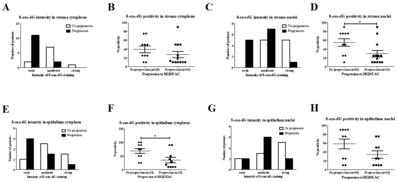
Figure 6.
(A–H): 8-oxo-dG staining in SIM progressors and non-progressors. (A) Chi-square test demonstrated significantly weaker intensity of 8-oxo-dG staining in patients with progressive disease (p = 0.008). (B) Mann–Whitney U test showed no difference in the percentage of stroma cytoplasm positive for 8-oxo-dG (p = 0.168). (C) Chi-square test showed weaker 8-oxo-dG staining in the progressors (p = 0.0201). (D) Non-progressive Barrett’s esophagus (BE) was associated with an increased percentage of positive stroma nuclei (p = 0.039). (E) Chi-square test showed no difference in staining intensity in epithelial cytoplasm (p = 0.174). (F) Epithelial cytoplasm percentage positivity was significantly higher in the non-progressing group (p = 0.030). (G) Chi-square test demonstrated no difference in 8oxo-dG intensity in epithelial nuclei between both groups (p = 0.320). (H) Mann–Whitney U test showed no difference in percentage positivity of 8-oxo-dG epithelial nuclei (p = 0.100). * p < 0.05.

