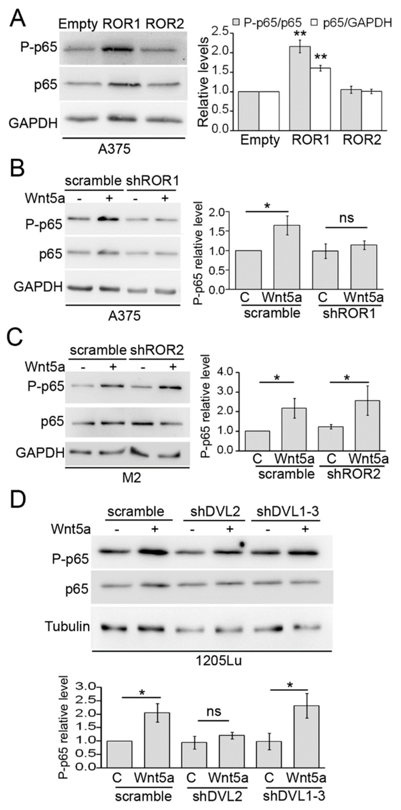
Figure 2.
ROR1 and Dvl2 are required for p65 phosphorylation by Wnt5a. (A) Western blot analysis of A375 cells transduced with either ROR1 or ROR2. (B) A375 cells were transduced with a ROR1 shRNA or a scrambled shRNA and stimulated with either Wnt5a or control media for 30 min. The proteins extracts were blotted with the indicated antibodies. (C) M2 cells were transduced with a ROR2 shRNA or a scrambled shRNA and stimulated with either Wnt5a or control media for 30 min. The proteins extracts were blotted with the indicated antibodies. (D) 1205Lu cells were transduced with lentivirus encoding, either Dvl2 shRNA or Dvl1-3 shRNA, or a scramble sequence as a control, and stimulated with either Wnt5a or control media for 30 min. The proteins extracts were blotted with the indicated antibodies. GAPDH or Tubulin was used as the loading controls. The blots displayed are representative of three independent experiments. The bar graphs show the mean ± SD (from three independent experiments) of P-p65 levels normalized to the level of total p65 detected after stripping the membrane. Results are expressed as the fold change relative to control-treated cells (lane 1 in all panels). The statistical significance was tested by a student’s t-Test (treated sample vs. paired control in B –D) or ANOVA (A), followed by Dunnett’s Multiple Comparison Test, using log transformed FC values. * p < 0.05, ** p < 0.01, n = 3.

