Figure 2.
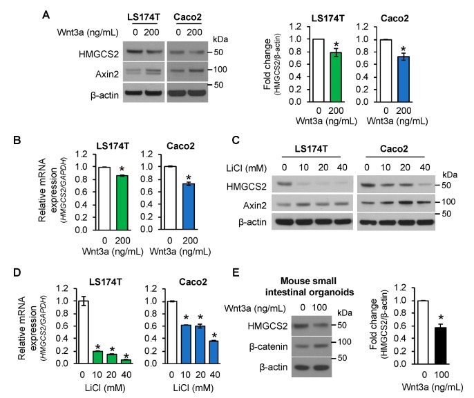
Activation of Wnt/β-catenin pathway suppressed HMGCS2 expression. (A,B) LS174T and Caco2 cells were treated with 200 ng/mL Wnt3a for 24 h. (A) Western blot analysis was performed using the antibodies as indicated. Densitometric quantification from three separate experiments was performed and is represented as fold change with respect to β-actin (n = 3, data represent mean ± SD; * p < 0.05 vs. 0 ng/mL Wnt3a). (B) The level of HMGCS2 mRNA was determined by real-time RT-PCR. (n = 3, data represent mean ± SD; * p < 0.05 vs. 0 ng/mL Wnt3a). (C,D) LS174T and Caco2 cells were treated with various dosages of LiCl or 40 mM of NaCl as control for 24 h. (C) Western blot analysis was performed using the antibodies as indicated. (D) The level of HMGCS2 mRNA was determined by real-time RT-PCR. (n = 3, data represent mean ± SD; * p < 0.05 vs. 40 mM NaCl). (E). Mouse small intestinal organoids were treated with 100 ng/mL Wnt3a for 3 days. HMGCS2 protein expression was determined by western blotting. Densitometric analysis from three independent experiments was performed and is represented as fold change with respect to β-actin (n = 3, data represent mean ± SD; * p < 0.05 vs. 0 ng/mL Wnt3a).

