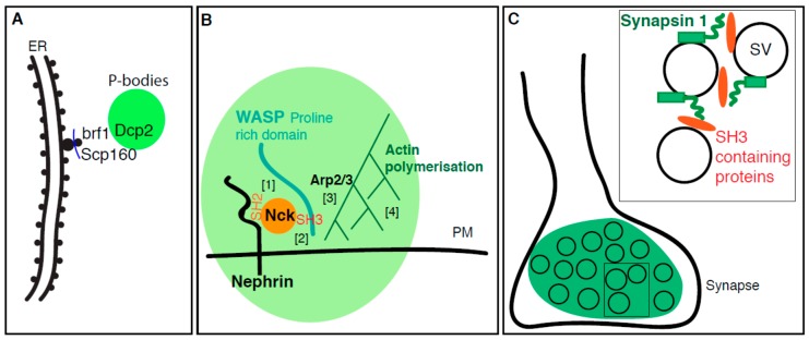
Figure 3.
Membrane enhances the formation of phase-separated condensates. (A): P-bodies form near the ER as DCP2 interacts with the ER proteins Scp160 and Brf1 themselves interacting with translating ribosomes present at the translocon. (B): Nephrin is a transmembrane adhesion receptor resident of the plasma membrane (PM). It binds to Nck-SH2 domain via its long disorder cytoplasmic tail [1]. Nck contains also three SH3 domain can binds the proline-rich domain of N-WASP [2]. This forms a biocondensation (green) that recruits Arp2/3 [3], resulting in actin polymerization around Nephrin [4] (C): Synapsin 1 at synapse interacts with several SH3 domain containing proteins (in orange such as Intersectin and Gbr2) via its intrinsically disordered region. These interactions are proposed to lead to the phase separation of these components and coalescence of synaptic vesicles (SV) as observed at the synapse.

