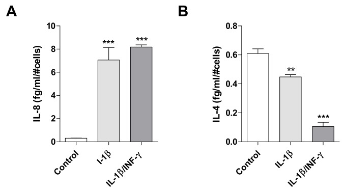
Figure 3.
Effects of inflammatory stimuli on release of IL-8 and IL-4 from C20 cells. The pro-inflammatory IL-8 (A) and anti-inflammatory IL-4 levels (B) were evaluated in conditioned medium derived by C20 cells treated with IL-1β (20 ng/mL) or IL1-β/INF-γ (100 ng/mL/ 50 ng/mL) for 24 h. The amount of interleukins measured was normalized to the number of cells and expressed as fg/mL/#cells. The bars represent the mean ± SEM of two different experiments performed in triplicate. The significance of the differences was determined by one-way ANOVA, followed by Bonferroni’s post-test: ** p ≤ 0.01, *** p ≤ 0.001, vs. control.

