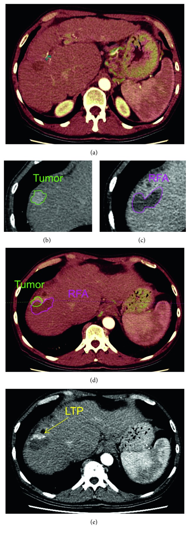
Figure 1.

Image analysis protocol. (a) Registration (overlay) of preinterventional and postinterventional CT scans. (b) Semiautomatic delineation of tumor volume. (c) Semiautomatic delineation of RFA volume. (d) Image fusion plane: margin analysis by overlaying pre- and postinterventional imaging. (e) Follow-up scan with local tumor progression.
