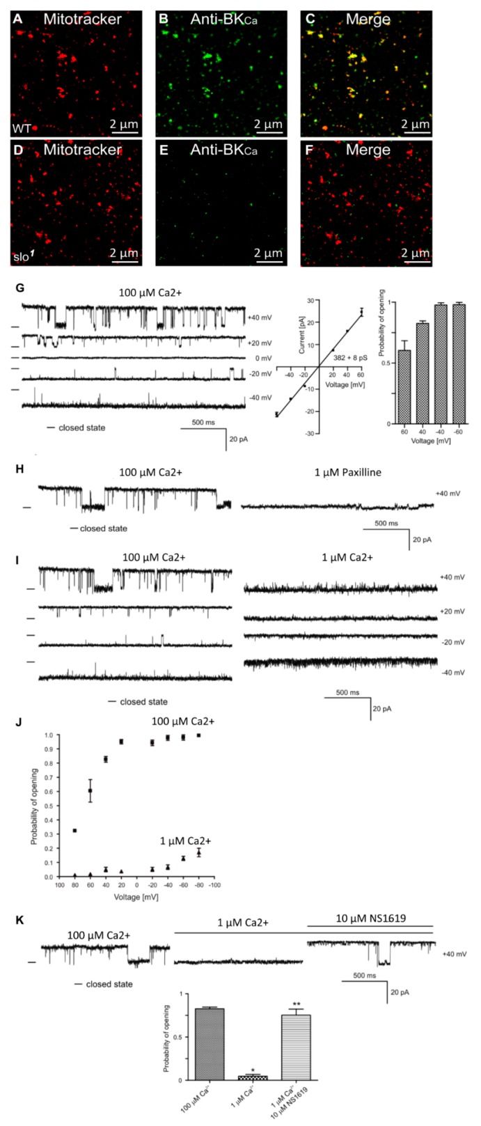
Figure 1.
Localization of dSlo in isolated mitochondria. High-resolution confocal images of isolated mitochondria from Drosophila (A–C, wild type, D–F slo mutants) loaded with mitotracker (A,D red) and labeled with an anti-Slo antibody (B,E green). Overlays are shown in (C,F). Protein proximity index for dSlo to mitotracker was 0.5 ± 0.1. (G), Single-channel current-time recordings (left panel), current-voltage characteristics (middle panel) and Po analysis of single-channel events in a symmetric 150/150 mM KCl isotonic solution (100 μM Ca2+) at different voltages in mitoplast prepared from whole flies. (H), Effects of 1 μM Paxilline on the single-channel activity. (I), Single-channel current-time recordings in symmetric 150/150 mM KCl isotonic solution at control (100 μM Ca2+) and after reduction calcium concentration to 1 μM Ca2+. (J), Analysis opening probability in the presence of 1 and 100 μM Ca2+ at different voltages of the mitoBKCa channel in mitoplast prepared from whole flies. All data were acquired in a symmetric 150/150 mM KCl isotonic solution (n = 4). (K), Current–time recordings of single-channel activity in symmetric 150/150 mM KCl isotonic solution at control (100 μM Ca2+), after reduction calcium concentration to 1 μM Ca2+ and after application of 10 μM NS1619. The bar graph shows the distribution of the Po under the conditions above. * p < 0.001 vs. the control. ** p < 0.001 vs. 1 μM Ca2+. The data in (G,J,K) are presented as the means ± S.D. The recordings were low-pass filtered at 1 kHz. “-“ indicate a closed state of the channel.

