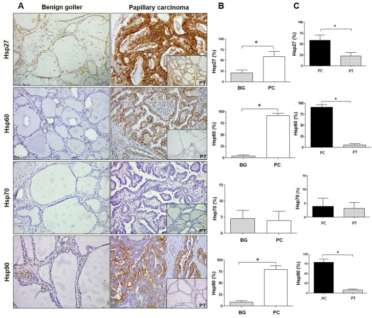
Figure 1.
Immunohistochemistry for Hsps in benign goiter and papillary carcinoma. (A) Immunohistochemistry images of Hsp27, Hsp60, Hsp70, and Hsp90 in human thyroid tissue of benign (non-toxic) goiter and papillary carcinoma with pertinent normal peritumoral tissue (PT; insets at bottom right of each panel on the right). Magnification 200×. (B) Histograms showing the percentage of immunopositivity for Hsp27, Hsp60, Hsp70, and Hsp90 in benign goiter (BG) and papillary carcinoma (PC). Data are presented as the mean ± SD. * p ≤ 0.0001. (C) Histograms showing the percentage of immunopositivity for Hsp27, Hsp60, Hsp70, and Hsp90 in samples of papillary carcinoma (PC) and normal peritumoral tissue (PT). Data are presented as the mean ± SD. * p ≤ 0.0001.

