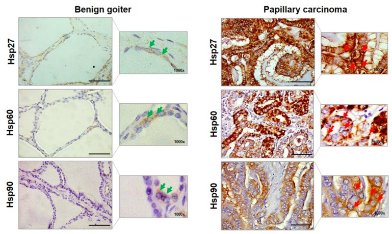
Figure 2.
Representative images of the immunohistochemistry of benign goiter and papillary carcinoma for Hsp27, Hsp60, and Hsp90. Larger images were acquired at a magnification of 400× (scale bar: 100 µm); smaller images at 1000× allowed a better visualization of the cellular localization of immunopositivity. Green arrows, in benign goiter images, indicate for Hsp27 the cytosolic and perinuclear localizations; for Hsp60 the cytosolic and cytoplasmic granular (i.e., mitochondrial) localizations; and for Hsp 90 the cytosolic localization. Red arrows, in papillary carcinoma, indicate the cytoplasmic and plasma–cell membrane (or close to this membrane) localizations of Hsp27; the cytoplasmic diffuse, close to, and in plasma–cell membrane immunopositivity of Hsp60; and cytosolic and plasma cell–membrane localizations of Hsp90.

