Figure 1.
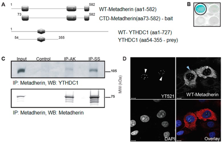
Metadherin interacts with YTHDC1 in the nucleus. (A) Schematic representation of metadherin (upper panel) and YTHDC1 constructs (lower panel). Lysine-rich domains (KRD) of metadherin shown in grey rectangles (top), and glutamine rich domain (QRD) of YTHDC1 shown in grey hexagon (bottom). (B) Yeast two-hybrid assay using pLexA-DIR-metadherin-aa73–582 (C-terminal domain (CTD)-metadherin) as bait and a human placental cDNA library. β-Galactosidase assay confirms interaction between YTHDC1 and metadherin, YTHDC1 well circled in black, adjacent wells show alternate targets which were all negative. (C) Endogenous metadherin was immunoprecipitated using two antibodies against a central epitope (IP-AK) and C-terminal epitope (IP-SS) with metadherin from 1 mg of COS7 protein lysate. Input is 10 µg protein lysate, sheep IgG was used as control. Western blots were probed for YTHDC1 and metadherin (SS). Blots were visualised with enhanced chemiluminescent luminol-based (ECL) substrate plus or, when the signal exceeded the dynamic range of film, using 3,3’-Diaminobenzidine (DAB). (D) COS7 cells were fixed in methanol, probed with an anti-YTHDC1 (red) and anti-metadherin (green) antibodies, and mounted with DAPI (blue). White arrows show nuclear localization of the target protein, blue arrows indicate cytoplasmic localization. Images were obtained using a Nikon Eclipse confocal microscope using a ×100 objective. Scale bars represent 10 μm.

