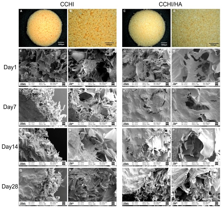
Figure 2.
Scanning electron microscopy images (E–T) of chitosan scaffolds with or without hyaluronic acid at different time points in chondrogenic medium culture, with less magnified overview (E,I,M,Q and G,K,O,S, respectively) and detail images in higher magnification (F,J,N,R and H,L,P,T, resp.). The sponge-like topography of non-cultured chitosan scaffold (A,B) and chitosan with hyaluronic acid scaffold (C,D) discs is shown before submersion into the medium. After 24 h in the chondrogenic medium with hTGF-β3 + hBMP-6, hADSCs were already well established and started to form a matrix (E–H). Human ADSCs in both scaffold types treated with the chondrogenic medium were observed to quickly and efficiently deposit substantial amounts of a fibrous matrix at Day 7 (I–L), filling up the microporous structures of the scaffolds. The matrix was aggregating into a woven fibrous structure by Day 14 (M–P). By Day 28, microstructures of the scaffold material could not be detected by SEM since the scaffolds were completely covered by ECM-like material (Q–T). Magnifications were set at 300× (E,G,I,K,M,O,Q,S), 1100× (F,H,J,L,N,P,R,T).

