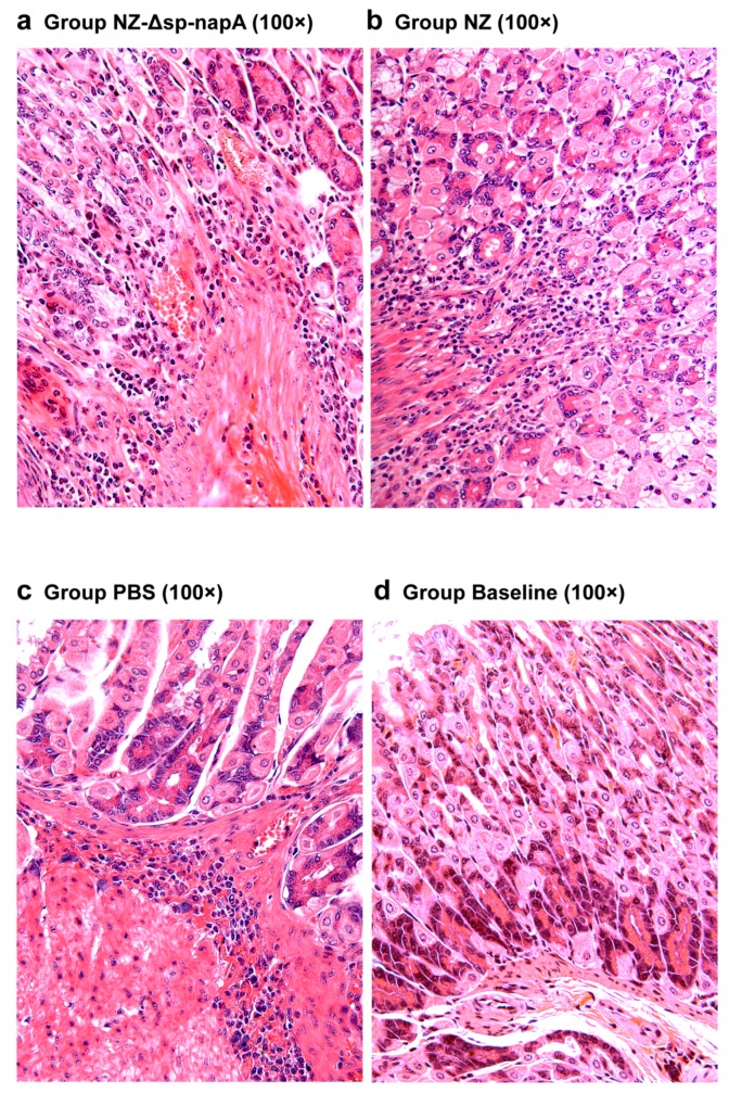
Figure 6.
Histological examination of gastric tissues of the LtB-untreated mice. NZ-Δsp-napA (a), NZ (b) and PBS (c) groups (n = 11 each) were gavaged with NZ3900/pNZ-Δsp-napA, NZ3900/pNZ8110 and PBS, respectively, prior to H. pylori challenges. No gavages were given to the Baseline group (n = 10) (d). The gastric tissues were sampled from the mice 1 week after the challenges and examined via paraffin section and hematoxylin-eosin staining. The mice of all the H. pylori-challenged groups had gastric inflammatory responses characterized by the infiltration of lymphocytes, neutrophils and microphages, abscess, and congestion in gastric mucosa and submucosa.

