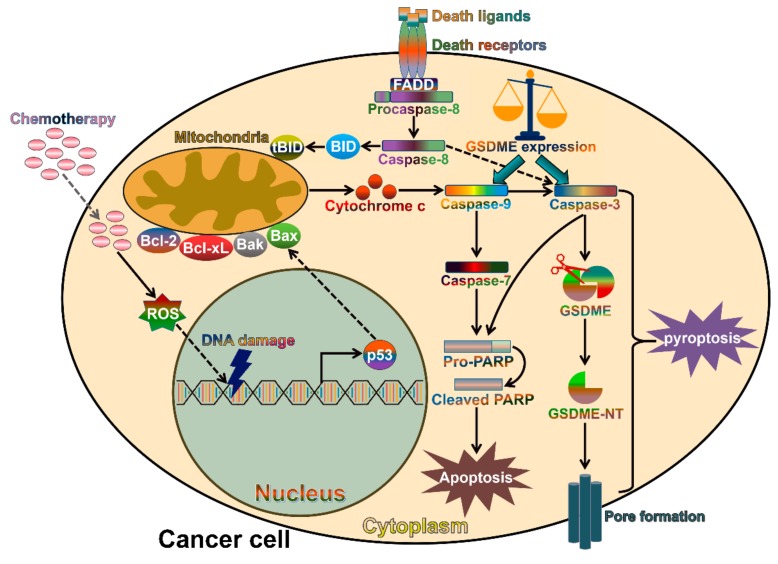
Figure 3.
Schematic representation of chemotherapy-induced apoptosis and pyroptosis in cancer cell. Apoptosis is induced via the intrinsic or extrinsic death signaling pathway. Chemotherapy induces DNA damage and activates p53, which interacts with Bax in mitochondria and triggers the mitochondria-dependent (intrinsic) apoptotic pathway. Detailedly, Bax promotes the release of cytochrome c and initiates the capase-9/caspase-3 cascade, resulting in cell apoptosis. In contrast, the extrinsic apoptosis pathway involves the binding of death ligands (e.g., Fas and TNF) to their receptors. This event gives rise to the activation of caspase-8. The latter evokes cell apoptosis by cleaving BID and directly activating caspase-3. In certain GSDME-expressing cancer cells, GSDME is able to switch chemotherapy-induced apoptosis to pyroptosis. Caspase-3 cleaves GSDME to generate the N-terminal fragment (GSDME-NT), which perforates the plasma membrane to execute pyroptosis. Bcl-2, B-cell lymphoma-2; Bcl-xL, B-cell lymphoma-extra large; Bak, Bcl-2 homologous antagonist/killer; Bax, Bcl-2-associated X protein; BID, Bcl-2 homology domain 3 (BH3)-interacting domain death agonist; tBID, truncated BID; FADD, Fas-associated death domain protein; ROS, reactive oxygen species; PARP, poly (ADP-ribose) polymerase; GSDME, gasdermin E; GSDME-NT, the N-terminal fragment of GSDME.

