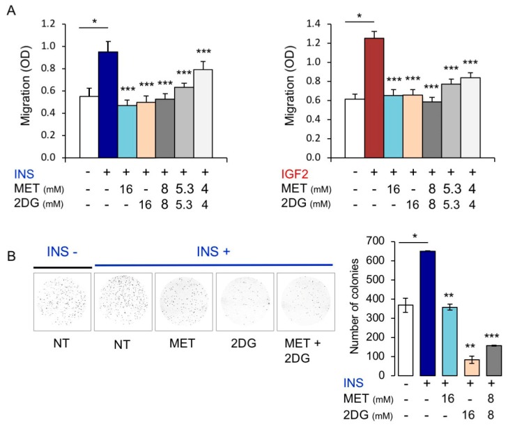
Figure 9.
Metabolic inhibitors negatively regulated insulin (INS) or IGF2-dependent biological responses of MCF7IGF1R-ve/IRA cells. (A) Cell invasion. Serum starved MCF7IGF1R-ve/IRA cells were seeded on polycarbonate filters coated with 25 µg/mL fibronectin and treated with either metformin (MET) or 2DG alone or in combination. Cells were then allowed to migrate for 6 h to the lower chamber in the absence (NT) or presence of 10 nM INS or IGF2. Values are means ± SEM of three independent experiments done in duplicate. Asterisks indicate statistical significance vs. INS or IGF2 treated condition in the absence of metabolic inhibitors (* p < 0.05; *** p < 0.001). (B) Colony formation. MCF7IGF1R-ve/IR-A cells were seeded in soft-agar and treated with either MET or 2DG alone or in combination, in the absence (NT) or presence of 10 nM INS (see Methods). Colonies were then stained with MTT and photographed. The histogram on the right represents the number of colonies (means and range). Data shown are from two independent experiments run in quadruplicate wells. Asterisks indicate statistical significance vs. INS treated condition in the absence of metabolic inhibitors. (* p < 0.05; ** p < 0.01; *** p < 0.001).

