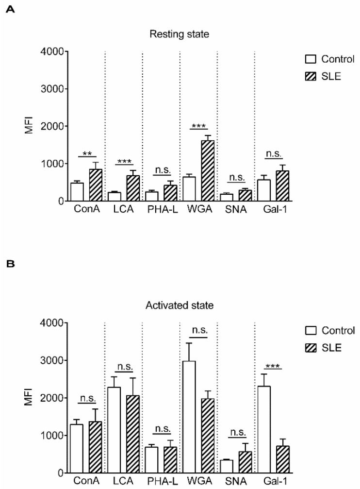
Figure 1.
Lectin binding properties of resting and activated T cells from healthy donors and from systemic lupus erythematosus (SLE) patients. Peripheral blood T cells were obtained from healthy controls and SLE patients. The cells were left unstimulated (resting state, A) or were activated with 1 µg/mL phytohaemagglutinin L (PHA L) for 72 h (activated state, B). Cells were stained with viability dye, fixed then labeled with anti-CD3-PE-Cy5 antibody, followed by fluorescein isothiocyanate (FITC)-conjugated lectin. The samples were evaluated with flow cytometry. Binding of FITC-conjugated lectins is shown as mean (±SEM) of the median fluorescence intensity (MFI) values of flow cytometry histograms of resting (A) or activated (B) CD3-positive live T cells. Lectin names are listed in Table 1. MFI: mean fluorescence intensity, ConA: concanavalin-A, LCA: Lens culinaris agglutinin, WGA: wheat germ agglutinin, PHA-L: Phaseolus vulgaris leukoagglutinin, SNA: Sambucus nigra agglutinin, Gal-1: galectin 1. Statistical analysis was performed using an unpaired Student t-test. ** p < 0.01; *** p < 0.001; n. s.: not significant. SLE: n = 18, and healthy controls: n = 19.

