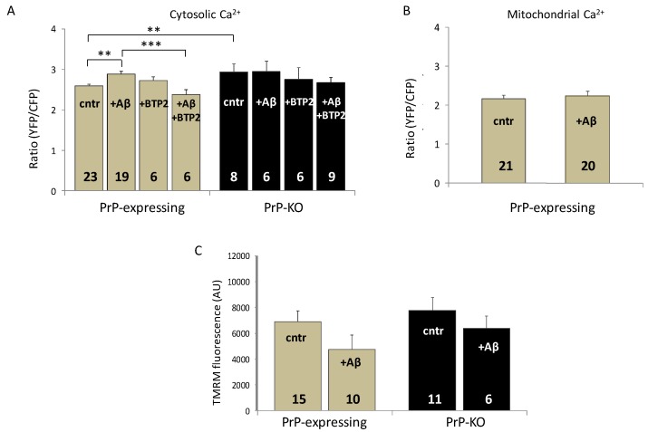
Figure 2.
Soluble Aβ1–42 oligomers abrogate the PrP-dependent regulation of resting cytosolic Ca2+ but does not affect mitochondrial Ca2+ levels and membrane potential in PrP-expressing primary cortical neurons. (A) Incubation with soluble Aβ1–42 oligomers (+ Aβ) increases resting cytosolic Ca2+ in PrP-expressing cortical neurons but not in PrP-KO neurons, nullifying the difference observed between the two untreated genotypes (cntr). While preincubation with the SOCE inhibitor N-{4-[3,5-bis(Trifluoromethyl)-1H-pyrazol-1-yl]phenyl}-4-methyl-1,2,3-thiadiazole-5-carboxamide (+ BTP2, 200 nM) leaves unaffected basal cytosolic Ca2+ in untreated cells, and it reverts the Aβ effect in PrP-expressing neurons (+ Aβ, + BTP2), upregulation of SOCE is the likely responsible for the Aβ-induced higher basal Ca2+ levels. (B) Conversely, Aβ does not affect Ca2+ levels in mitochondria of PrP-expressing neurons. Bar diagrams of panels A and B report the ratio between the FRET-acceptor (yellow fluorescent protein, YFP) and -donor (cyan fluorescent protein, CFP) fluorescence intensities of the used cameleon Ca2+ probe (D1cpv and 4mtD3cpv [71] in the cytosol and in the mitochondrial matrix, respectively). For these measurements, four days after isolation and plating, neurons were transduced with adeno-associated virus (AAV) coding for the chameleon bearing no targeting sequence (A), or a sequence directing the probe to the mitochondrial matrix (B), under the control of the pan-neuronal hSyn promoter [75]. Two days after infection, neurons were mounted into an open-topped chamber and maintained in 1 mM CaCl2-containing Krebs-Ringer buffer kept at 37 °C using a temperature-controlled apparatus (TC-324B, Warner Instruments). FRET measurements and analysis were performed as previously reported [75]. (C) No statistical variation is observed between the mitochondrial membrane potential of untreated, or Aβ-treated, PrP-expressing, and PrP-KO neurons. Here, the bar diagram reports the relative fluorescence quantification of the membrane potential-sensitive probe tetramethylrhodamine methyl ester (TMRM, Molecular Probes) [46] in experiments carried out with neurons loaded with TMRM (20 nM, 30 min, 37 °C) in the presence (+Aβ), or in the absence (cntr), of Aβ1-42 oligomers. ** p < 0.01, *** p < 0.001 (Student′s t-test). AU (arbitrary units). Other details are as in the legend to Figure 1.

