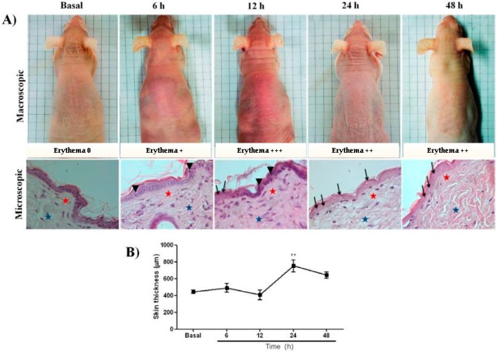
Figure 1.
(A) Macro- and microscopic changes induced by UVB in skin of hairless mouse. Dorsal skin was exposed to a single UVB dose (2.4 J cm−1) and changes were monitored at various intervals. Skin biopsies were kept in 10% buffered formalin and processed for histopathological examination. Representative photomicrographs using 40× objective of Haematoxylin-eosin at 6, 12, 24, and 48 h post UV were compared with basal group. Apoptotic cell = arrowhead; enucleated cells = arrows; superficial dermis = red stars; deep dermis = blue stars). (B) Morphometric analysis of basal, 6, 12, 24, and 48 h after irradiation. The data represent total skin thickness (10× objective): mean ± SEM of 6–8 animals in each group (** p < 0.01 difference versus basal, ANOVA, Newman–Keuls post-test).

