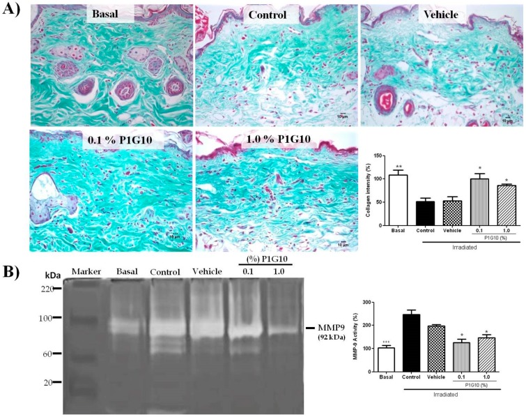
Figure 7.
Changes in collagen and MMP-9 activity induced by P1G10 after UVB-irradiation. Mice were exposed to UVB (2.4 J cm−1) and treated with Natrosol gel (vehicle) or 0.1% and 1% P1G10 for 24 h post-irradiation. Biopsies were preserved in 10% buffered formalin for histological analysis. (A) Photomicrographs illustrate collagen fibers from dermis in slides stained with Gomori trichrome (40× objective). The graph represents the area corresponding to collagen fibers obtained from the image after digital processing. (B) MMP-9 activity was assessed by zymography and data expressed as percentage MMP-9 activity relative to basal value (100%). Results represent the mean ± SEM of 6–8 animals per group (* p < 0.05, ** p < 0.01 and *** p < 0.001 difference versus vehicle, ANOVA, Newman–Keuls post-test).

