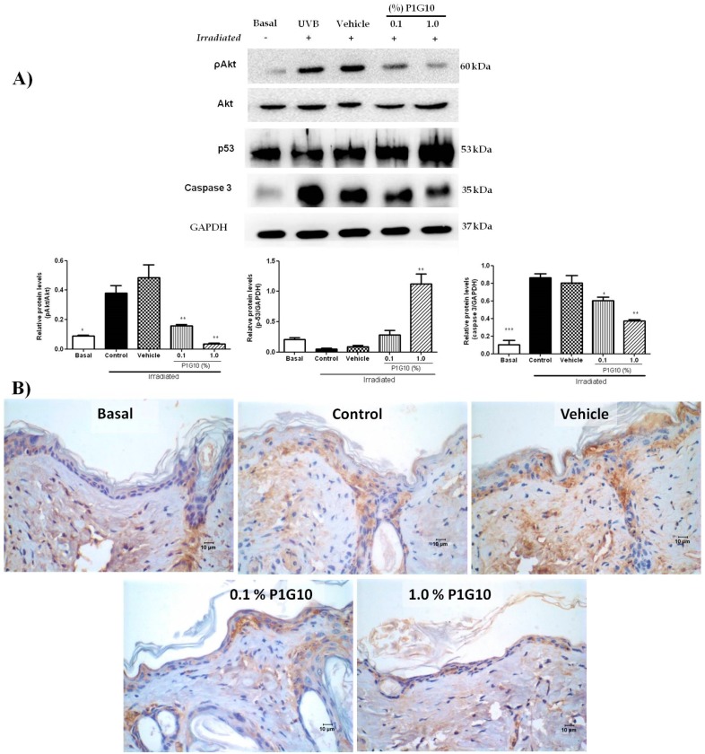
Figure 9.
P1G10 alters the expression of pakt, p53, and caspase-3 in irradiated skin. Mice were exposed to UVB (2.4 J cm−1) and treated with Natrosol gel (vehicle) or 0.1% and 1% P1G10. After 24 h, the skin of euthanized animals was dissected and processed for analysis. Cell lysates were used to assess pakt, p53, and caspase-3 (A). The data show a western blot and densitometric analysis. Protein levels were normalized to GAPDH using ImageQuant software. (B) A brown color indicates the presence of caspase-3 positive cells in irradiated samples. Images captured with 40× objective, Scale = 10 µm. Results represent the mean ± SEM of 6–8 animals per group (* p < 0.05, ** p < 0.01 and *** p < 0.001 difference versus vehicle, ANOVA, Newman–Keuls post-test).

