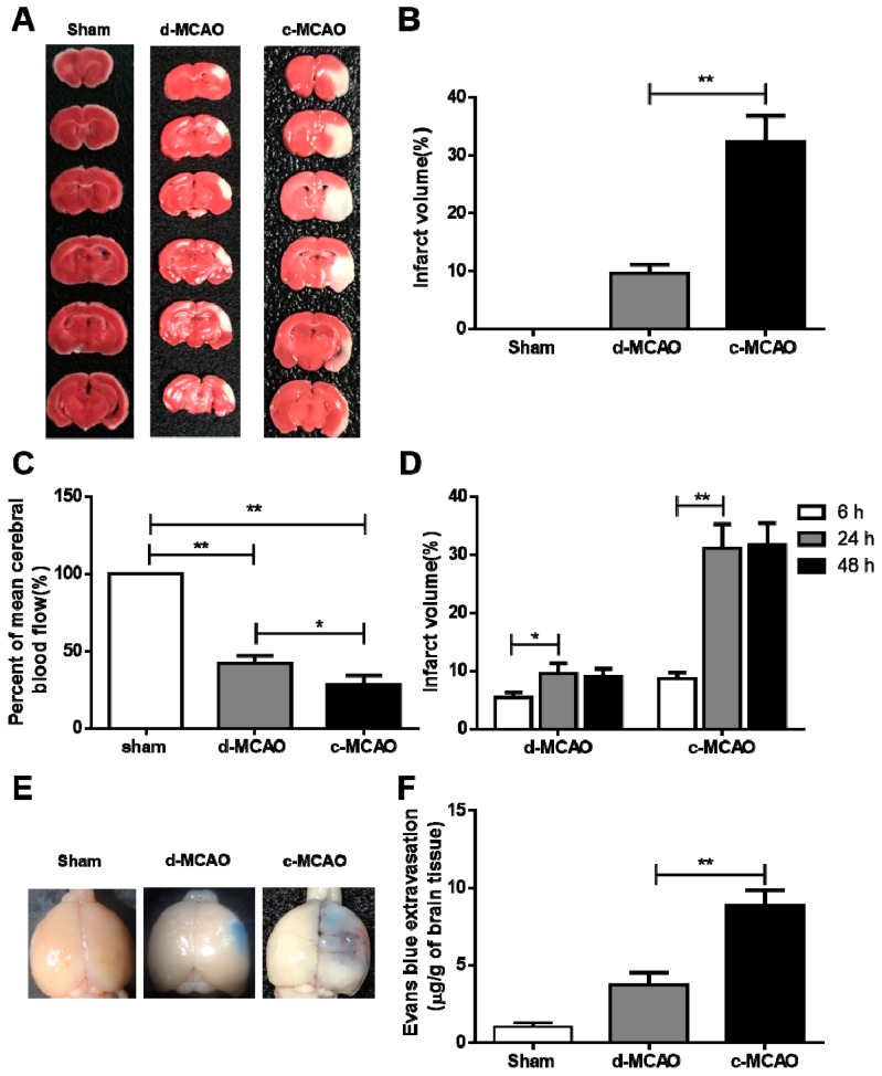
Figure 2.
Comparison of the severity of ischemic strokes induced by d-MCAO and c-MCAO. Wild-type (WT) hamsters were subjected to d-MCAO and c-MCAO for 24 h.(A) Representative images of six coronal brain sections stained with 2, 3, 5-triphenyltetrazolium chloride (TTC) staining. (B) Quantification of the infarct volumes in Sham, d-MCAO, and c-MCAO from (A) (n = 6/group). (C) Measurement of cerebral blood flow after ischemia by laser Doppler transducer (n = 6–8/group). (D) Time course analysis of infarct volume in ischemic hamsters induced by d-MCAO and c-MCAO (n = 6–8/group). (E) Representative images of the Evan’s blue (EB)-stained whole brains in sham, d-MCAO, and c-MCAO group. (F) Quantification of the Evan’s blue extravasation; data are expressed as mean ± SD. * p < 0.05; ** p < 0.01 versus sham group.

