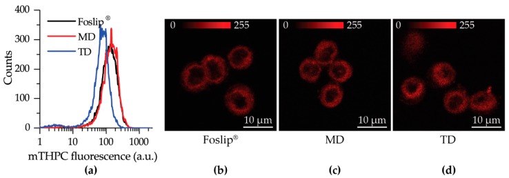
Figure 4.
(a) Flow cytometry histograms of HT29 monolayer cells treated with Foslip® (black), MD, (red) and TD (blue) for 24 h; (b–d) Typical confocal images of mTHPC fluorescence in HT29 monolayer cells at 3 h post-incubation with (b) Foslip®, (c) MD, and (d) TD. Scale bar: 10 μm. The concentration of mTHPC was 1.5 μM. Serum concentration was 2%.

