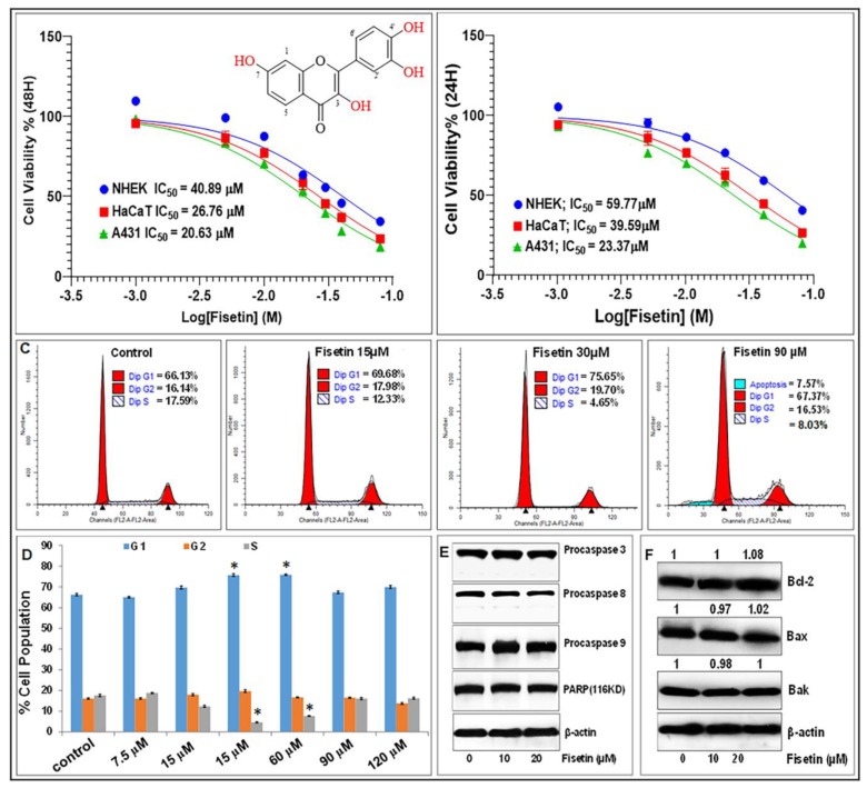
Figure 1.
Fisetin at low doses (< 20 µM) does not significantly affect primary normal human epidermal keratinocyte (NHEK) growth and viability and does not induce apoptosis. (A/B) Relative number of viable NHEK, immortalized keratinocytes (HaCaT), and A431 cancer cells treated with or without fisetin (1–80 µM) for 24 h (A) and 48 h as determined by MTT assay. Mean of percentage viability of NHEK, HaCaT, and A431 cell lines plotted against the indicated doses of fisetin. Experiments were performed three times with each concentration done in octuplicate wells, and the IC50 values calculated from these plots are shown. (C) Effects of fisetin on the percentage of cells population in the different phases of cell cycle, and indication of late apoptosis or necrosis (i.e., PI-positive) cells were only seen in cells treated with higher fisetin concentration. (D) Levels or percentahes of cell cycle distribution in fisetin-treated cells as assessed by flow cytometry analysis. Bar graphs represent mean ± SD of results from three independent experiments performed in triplicate. Statistical significance was determined using one-way ANOVA and Dunn’s multiple comparison test and significance was considered when p < 0.05 (*), as compared with the control. (E) and (F) Effect of different concentrations of fisetin on the expression of markers of apoptosis including caspase-3, -8, and -9, PARP (85 kDa and 116 kDa) and Bcl-2 family of proteins (Bcl2, Bax, and Bak) on cells harvested after 48 h of treatment as analyzed by Western blotting. Equal protein loading was confirmed using β-actin as loading control. (F) Numerical data above the blots represent relative quantitative density values for the blots normalized with an internal loading control. The Western blot data shown are representative immunoblots of two to three independent experiments with similar results.

