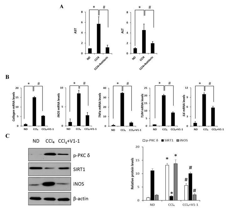
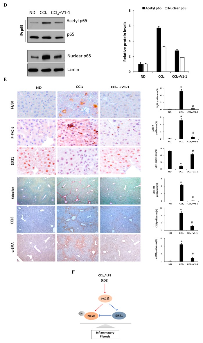
Figure 5.
PKCδ peptide inhibitor attenuates the progression of CCl4-induced liver fibrosis in mice. (A) Serum aspartate transaminase (AST) and alanine transaminase (ALT) levels from the control, CCl4, and CCl4 + V1-1 group mice. (B) The mRNA levels of collagen, iNOS, TNF-α, TLR4, and IL-6 in liver tissue homogenates were measured by qRT-PCR. Each value is the mean ± SD of three experiments. (C) The expression levels of p-PKCδ, SIRT1, and iNOS in the liver tissue were measured by immunoblotting analysis. (D) Liver tissue lysates were subjected to immunoprecipitation with anti-p65 antibodies. Immunoblot analysis was then used to detect the levels of acetyl p65 and total p65 in the immunoprecipitated complexes with anti-acetyl-lysine and anti-p65 antibodies, respectively (upper panel). The lower panel represents immunoblot analysis to detect nuclear p65 levels in liver tissue lysates without immunoprecipitation. (E) Representative liver tissue sections from ND, CCl4, and V1-1 peptide-treated CCl4 mice were subjected to immunohistochemical analysis for F4/80, phosphorylated PKCδ, and SIRT1 (upper panel group; original magnification, 200×) and Sirius Red, CK18, and α-SMA (lower panel; original magnification, 100×). * p < 0.05 compared to the control and # p < 0.05 compared to the CCl4 group. (F) The proposed signaling pathway for liver fibrosis where PKCδ upregulates NF-κB signaling but downregulates SIRT1 signaling simultaneously. In addition, SIRT1 directly downregulates NF-κB through deacetylation.(Red arrow: activation and Blue bar: inhibition).


