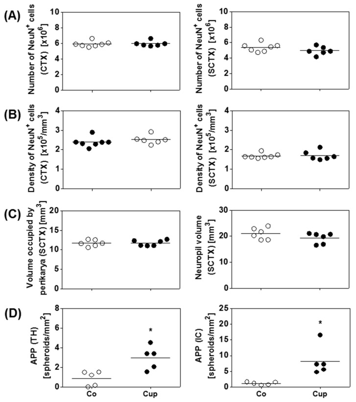
Figure 4.
Effects of chronic demyelination on the neuro-axonal/-dendritic compartment. (A) Numbers and (B) densities of neurons in the cerebral cortex (CTX) and the subcortical area (SCTX) of control (Co) (n = 6) and 12 weeks of cuprizone-intoxicated (Cup) mice (n = 6) analyzed with design-based stereology. (C) Neuropil volumes and volumes occupied by nerve cell perikarya in the SCTX of Co (n = 6) and 12-week Cup mice (n = 6). (D) Quantification of amyloid precursor protein (APP)-positive spheroids in the thalamus (TH) and internal capsule (IC) of chronically demyelinated mice (n = 5). Data are shown as individual values and means per group (lines). Statistical analysis revealed significant differences between Co and Cup in (D) (TH, p = 0.02; IC, p = 0.02); *p < 0.05.

