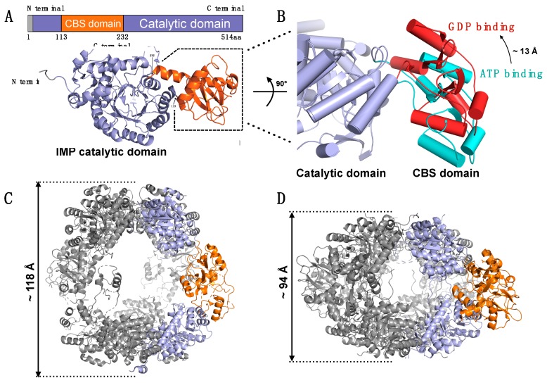
Figure 9.
Schematics of IMPDH structures. (A) Schematic representation of the human IMPDH2 protein (upper) and monomer of human IMPDH2 structure (lower) (PDB ID: 6i0m). IMPDH structure is shown in cartoons while α-helixes are shown in a coiled model. The CBS domain is colored orange, and the MPDH catalytic domain is shown in light purple. (B) Structural changes of the CBS domain upon ATP (Cyan) and GDP (Red) binding in monomeric IMPDH from Ashbya gossypii. Superposed ATP binding (PDB ID: 5mcp) and GDP binding (PDB ID: 4z87) by using Cα overlap and 243 aa was aligned. A-helixes were shown in a cylindrical model. The CBS domain rotated toward an IMPDH catalytic domain (light blue) significantly when GTP binds to the CBS domain, compared with ATP binding. (C,D) Different octameric forms between ATP binding (C) and GTP binding (D). Two monomers of octameric IMPDH (Gray) are colored orange (CBS domain) and light purple (catalytic domain). The approximate longitudinal dimensions of the octamers are indicated on their side. Comparing to ATP binding (C), the interaction changes between CBS domains upon GTP binding made the octameric structure of human IMPDH2 (D, PDB ID: 6i0o) more compact. Since no ATP-bound structure of human IMPDH has been determined, IMPDH from Ashbya gossypii (PDB ID: 5MCP) is used as the ATP binding model.

