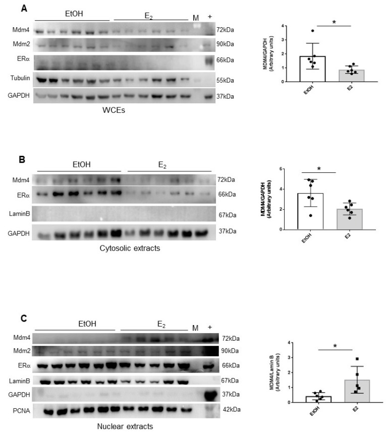
Figure 4.
E2 alters the subcellular localization of Mdm4 in mouse thymocytes. (A–C), WB analysis of whole cell extracts (WCEs) (A) cytosolic extracts (B) and nuclear extracts (C) of thymocytes of Mdm4Tg15 females treated with a single i.p. dose of physiologic solution or 50 µg/Kg E2 and sacrificed after 16 h. Laminin and GAPDH were used as LC. Graphs show mean ± SD of MDM4 levels quantified by Alliance V_1607 software (two-tailed unpaired t-test, WCEs: t = 2.443, df = 10, * p = 0.0346, Cytosolic extracts: t = 2.629, df = 10, * p = 0.0252, Nuclear extracts: t = 2.891, df = 9, * p = 0,0179).

