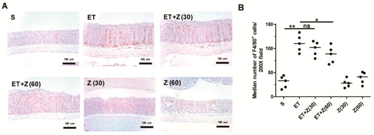
Figure 4.
Histological analysis of macrophage infiltration in distal colon of ETBF-infected mice. FFPE distal colonic tissues obtained from mice at day 7 post-infection were stained with anti-F4/80 antibody and counterstained with hematoxylin. Representative images are shown. S, sham; ET, ETBF; Z (30), Zerumbone (30 mg/kg); Z (60), Zerumbone (60 mg/kg). (A) Immunohistochemistry (IHC) for F4/80+ cells, ×200 magnification; Scale bar, 100 μm. (B) Median number of F4/80+ cells at 200× field. Scatter plot. Horizontal bar, median. * p < 0.05, ** p < 0.01. ns, no statistical significance.

