Figure 1.
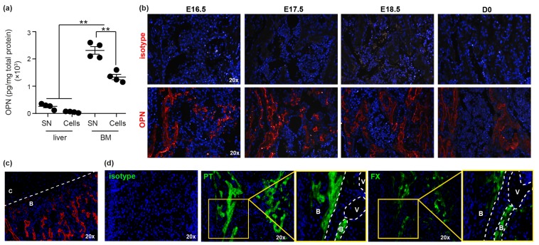
Osteopontin (OPN) is highly expressed in fetal BM. (a) OPN protein in E17.5 fetal liver and bone marrow (BM) was quantified using an OPN ELISA (R&D; MOST00). SN: supernatant. ** p < 0.01. (b) Immunohistochemical analysis of mouse E16.5, E17.5, E18.5 and D0 BM stained with either isotype control or anti-OPN (red). Grey areas represent autofluorescence. (c) E17.5 BM demonstrating lack of OPN expression in growth plate cartilage (C) compared to bone (B). (d) Immunohistochemical analysis of mouse E17.5 BM stained with either isotype control or anti-prothrombin (PT) and anti-factor X (FX). White dotted lines delineate the structures of the fetal femurs. B: bone; V: blood vessel; C: cartilage.

