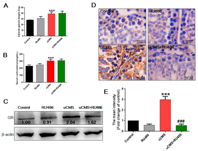
Figure 6.
Stress activated the HPA axis and elevated GR levels in the testes. (A) uCMS exposure increased the weight of the adrenal glands (n = 10). (B) uCMS exposure increased the serum corticosterone level (n = 10). (C) The protein levels of GR in testes were detected by Western blotting (n = 10). (D) Immunohistochemistry was used to detect the in situ protein levels of GR in testes (n = 10,400× magnification). White arrows indicated the GR-positive cells in testes. (E) Immunoreactivity intensity of GR was analyzed using Image Pro Plus6.0 software. Data were analyzed by one-way ANOVA with post hoc multiple comparisons test. * p < 0.05, ** p < 0.01, and *** p < 0.001 compared with the control group; #p < 0.05, ##p < 0.01, and ###p < 0.001 compared with the uCMS group. HPA = Hypothalamic–pituitary–adrenal; GR = glucocorticoid receptor; uCMS = unpredictable chronic mild stress; ANOVA = analysis of variance.

