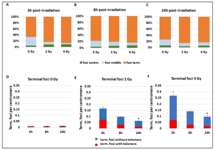
Figure 5.
Localization of γ-H2AX foci on chromosomes. Blood samples from three donors were irradiated at 2 or 4 Gy or kept free from ionising radiation and then analysed using the PCC technique. (A–C) After immunofluorescence staining, γ-H2AX foci position on chromosome sequence was determined “close to chromosome extremities”, “close to centromere”, or “in the middle of the chromatide”. Analyses were performed at 3 h (A), 8 h (B), or 24 h (C) after radiation exposure. (D–F) Only γ-H2AX foci “close to chromosome extremities” were sub-classed in two groups, γ-H2AX foci associated “with telomeres” or “without telomere” and followed at 3 h, 8 h, and 24 h post radiation exposure. The graphs show the repartition of foci without irradiation (D) and after 2 Gy exposure (E) or 4 Gy (F) radiation exposition. The decrease of terminal foci without telomere is significant at 24 h (p < 0.05).

