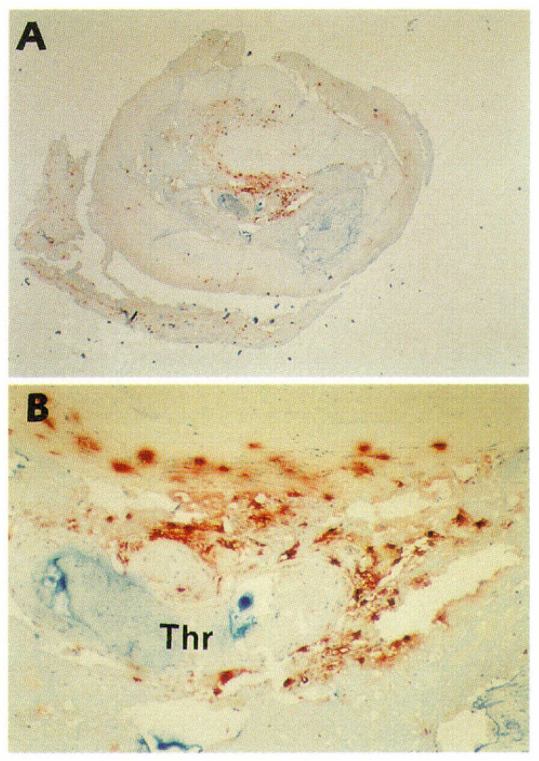
Figure 1.
Mast cells in an advanced atherosclerotic plaque of a highly atherosclerotic human coronary artery. This is a histological cross section of an eroded coronary segment with thrombotic occlusion. The specimen was from a 78-year-old man who died of myocardial infarction 3 h after the onset of symptoms of an acute coronary event. Mast cells were stained with monoclonal antibody G3 for the mast cell-specific neutral serine protease tryptase (red-brown). (A) Original magnification ×20. (B) Detail of the erosion site; original magnification × 200. “Thr” indicates thrombus. Numerous macrophages and T lymphocytes also accumulated in this area (not stained in this particular section). Reproduced from Kovanen PT, Kaartinen M, Paavonen T. Infiltrates of activated mast cells at the site of coronary atheromatous erosion or rupture in myocardial infarction [57].

