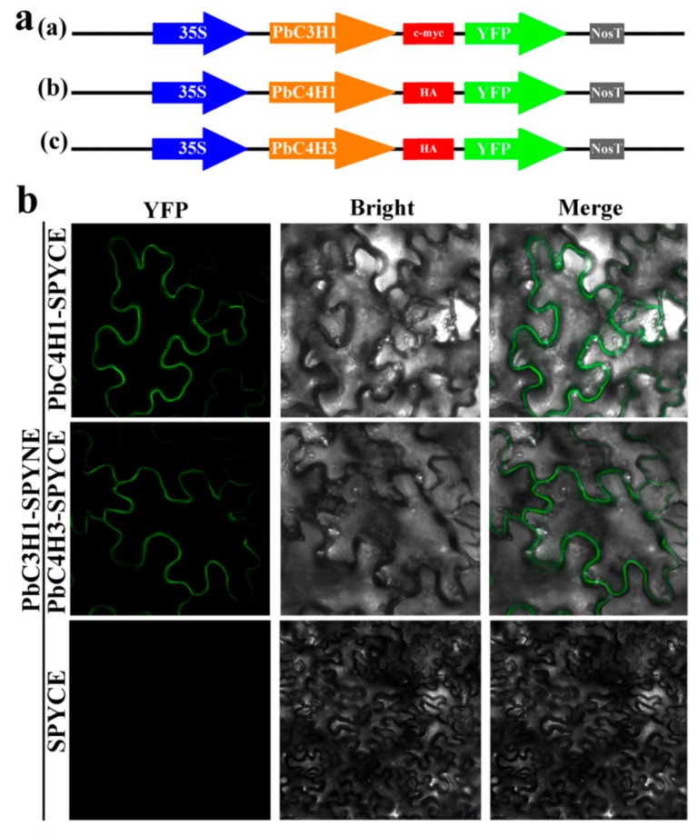
Figure 13.
In vivo BiFC analysis of interaction between PbC3H and PbC4H co-expressed in N. benthamiana leaf cells. The coding regions of PbC3H and PbC4H were fused to the N-terminal and C-terminal halves of YFP, respectively. (a): Schematic representation of the 35S: PbC3H1-YFP, 35S: PbC4H1-YFP, and 35S: PbC4H3-YFP fusion constructs used for transient expression. (b): A laser confocal microscope (Zeiss LSM700, Germany) was used to capture the fluorescence signals of the reconstituted YFP of the lower epidermal cells of leaves cut four days injection.

