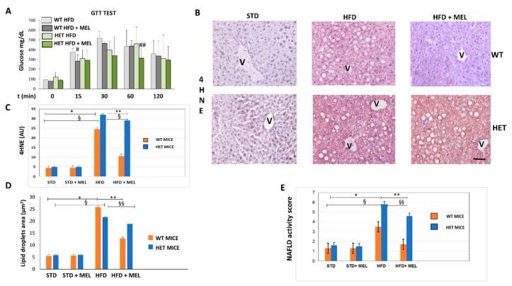
Figure 1.
Melatonin attenuated glycaemia, lipid peroxidation and steatosis in the liver of wild type (WT) mice, not in SIRT1 heterozygous (HET) mice placed on a high-fat diet. (A) Glucose transport test (GTT) measured blood glucose in WT and HET mice fed a maintenance (STD) or high-fat diet (HFD) for 16 weeks. # p < 0.05 vs. WT HFD at 15 min; ## p < 0.05 vs. HET HFD at 60 min. (B) Representative pictures of 4-hydroxynonenal (4HNE) immunostaining labeled as brown color drop. Intense lobular signal was evident in hepatocytes in HFD mice but melatonin reduced 4HNE staining in WT, not in HET. (C) The quantification of 4HNE immunohistochemistry. (D) Lipid droplets area estimated on semithin sections. (E) Non-alcoholic fatty liver disease (NAFLD) activity score an index of liver damage. STD: standard rodent diet; HFD: high-fat diet; HFD + MEL: high fat diet plus melatonin; V: central vein; scale bars indicate 10 µm in A and B. Values are mean ± SEM. * p < 0.05 vs. WT STD, ** p < 0.05 vs. WT HFD, § p < 0.05 vs. HET STD, §§ p < 0.05 vs. HET HFD.

