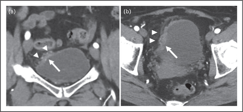
FIGURE 1.

A 63-year-old woman presented with hematuria. (a) A focal area of wall thickening at the dome of the bladder was shown on coronal CT postcontrast image of the pelvis (arrow). (a,b) Areas of fat stranding and nodularity adjacent to focal wall thickening (arrowheads) were highly suspicious for invasion of bladder wall. TURBT disease showed invasion only of the lamina propria. CT, computed tomography; TURBT, transurethral resection of the bladder tumor.
