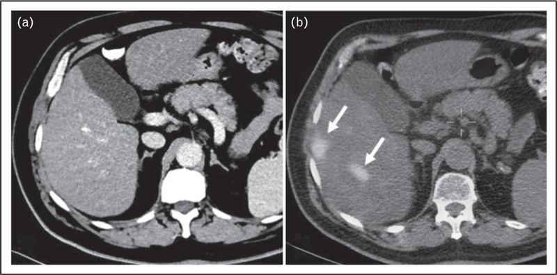
FIGURE 2.

A 65-year-old man with MIBC underwent screening CT examination of chest, abdomen, and pelvis (CAP) with contrast. (a) CT showed no evidence of metastasis to the liver. (b) PET/CT showed foci of increased radiotracer uptake within segments V and VI of the liver, with SUVmax of 9.2 compatible with metastases (arrows). Restaging follow-up CT CAP 8 weeks after this study confirmed metastasis to the liver. CT, computed tomography; MIBC, muscle-invasive bladder cancer.
