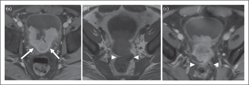
FIGURE 3.

A 37-year-old man presented with a large enhancing mass within the left body of the bladder. (a) Contrastenhanced pelvic MRI showed fat suppression extending into the trigon (arrows). (b) There is obliteration of the fat plane between bladder and rectum on nonfat-saturated T1-weighted images (arrowheads) and (c) Postcontrast T1-weighted images compatible with extravesical extension of the bladder mass and invasion of the rectum.
