
Fig. 2—
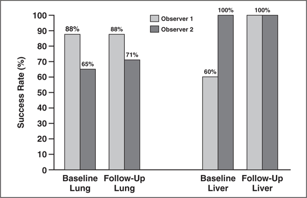
Graph shows percentage segmentation with lesion management application for lung and liver lesions on CT images from baseline and follow-up examinations. For lung lesions (n = 17) on baseline, the success rates were 88% for observer 1 and 65% for observer 2. For follow-up lung lesions, success was 88% for observer 1 and 71% for observer 2. For baseline liver lesions (n = 5), 60% were successfully segmented by observer 1 and 100% for observer 2. On follow-up liver segmentations, all lesions were successfully segmented for both observers.