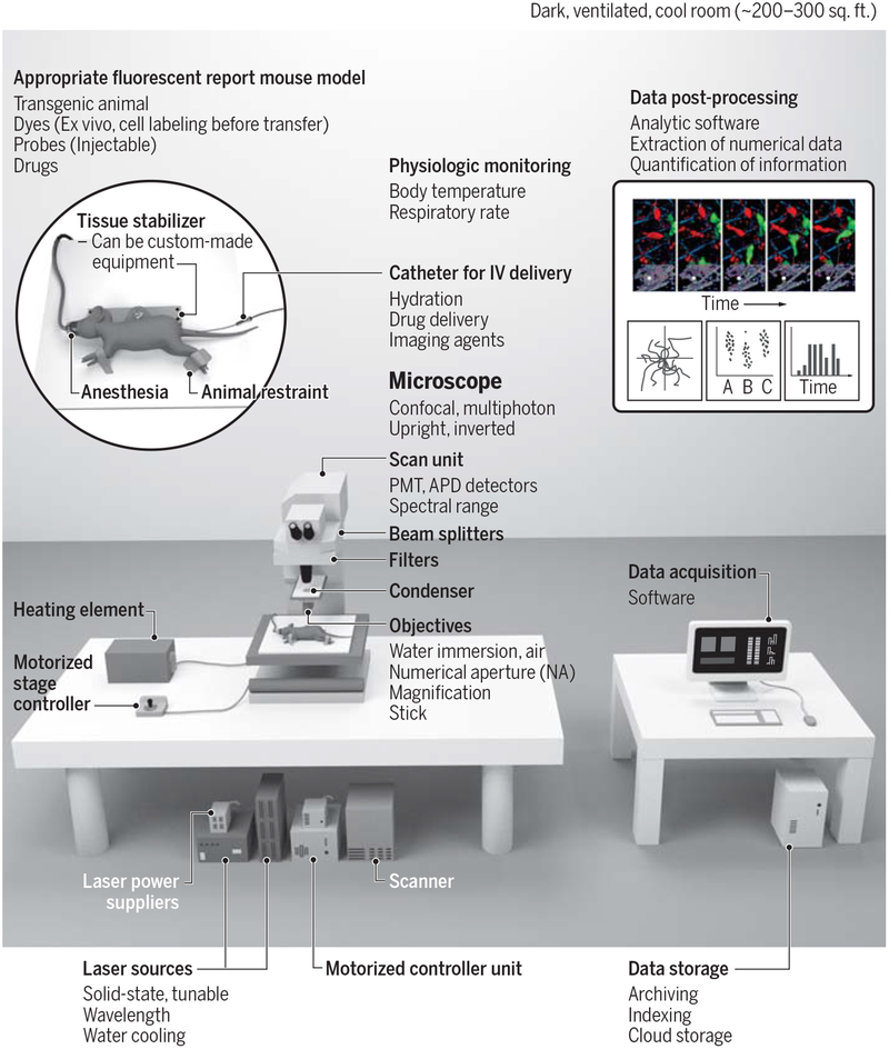
Fig. 1 – Diagram of an intravital imaging setup.
Left: the equipment required for conducting an intravital imaging experiment includes an appropriate fluorescent mouse model, a microscope (shown here is an upright multiphoton microscope), laser sources, physiological monitoring, an appropriate anesthesia setup, and a tissue stabilizer to prevent motion artifacts during imaging. This image illustrates imaging in a dorsal skinfold window chamber; other tissues can be imaged (see Figure 2), each of them requiring their own methods of tissue preparation, stabilization and monitoring. Right: data acquisition, followed by data storage and data post-processing to extract quantitative information. Credit: A. Kitterman/AAAS

