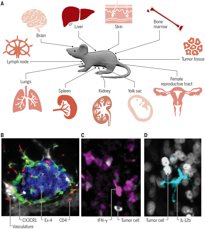
Fig. 2 – Existing applications of immune cell imaging in various tissues.
(A) A wide range of mouse tissue sites is adapted for single cell imaging. (B) Intravital micrograph showcasing detection of two immune cell types, namely CD4+ T cells (red) and CX3CR1+ macrophages (green) in pancreatic islets. β-cells (blue) were visualized with exendin-4–like neopeptide conjugated to the fluorescent dye Se-Tau-647 and the vasculature was detected with 500 K MW dextran conjugated to a Pacific blue dye (grey). (C-D) Intravital micrographs showcasing detection of cytokines produced by immune cells in live mice, here in tumor tissues. Panel C shows IFN-γ-producing lymphocytes (magenta) and panel D shows IL-12b-producing myeloid cells (cyan). Tumor cells are also shown (grey). All scale bars represent 10 μm. Credit: A. Kitterman/AAAS

