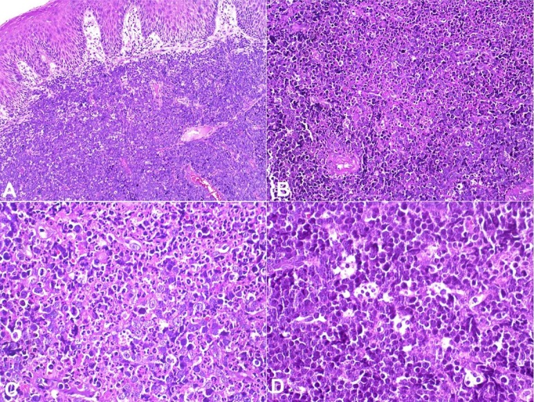
Figure 2. Histopathological features of sporadic Burkitt lymphoma. A, B – diffuse proliferation of lymphoid cells, with scarce cytoplasm, and round nuclei (H&E, A:10x, B:20x); C – The lymphoid cells are medium sized with multiple and single evident nucleoli, nuclear pleomorphism, numerous mitotic and apoptotic figures (H&E, 40x); D – The tumor cells and tingible body macrophages showing a “starry sky” pattern; the lymphoid cells show fine, granular and dense chromatin (H&E, 40x).

