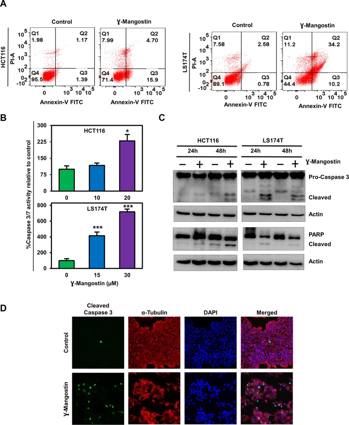
Figure 2. γ-Mangostin induces apoptosis.
(A) Cells were incubated with γ-Mangostin for 48h and assessed for apoptosis by Annexin V/PI staining. The following are the representations, Q1: Necrosis, Q2: Late apoptosis, Q3: Early apoptosis, Q4: Live cells. γ-Mangostin treatment induces apoptosis in HCT116 and LS174T cells. (B) Cells were incubated with γ-Mangostin for 24 h and assessed for apoptosis by Caspase3/7 assay. γ-Mangostin treatment results in significant increase in Caspase3/7 activity in HCT116 and LS174T cells (*p<0.05 and ***p<0.001). (C) γ-Mangostin treated HCT116 (10 µM) and LS174T (15 µM) cell lysates were analyzed for Caspase 3 and PARP expression. Both the cell lines showed cleaved Caspase 3 and cleaved PARP expression after γ-Mangostin treatment. (D) Immunofluorescence of HCT116 cells, untreated (top) and treated with 10 µM γ-Mangostin (bottom) were evaluated for Cleaved Caspase 3 (green), α-Tubulin (Red) staining with DAPI mounting. Treatment induces increased cleaved caspase 3 expression in γ-Mangostin treatment.

