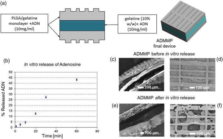
Figure 1.

(a) Scheme of ADMMP preparation; (b) release trend of adenosine from the sterilised final scaffold with adenosine loading (ADMMP). SEM images of section and surface of ADMMP (c, d) before and (e, f) after release at 24 hr. The section image of ADMMP before release test shows the thickness of the two microstructured external layers and the thickness of the internal layer containing gelatin and adenosine. The section image after 24‐hr release in MilliQ water points out an intermediate empty space. The surface image of the scaffold shows the presence of free voids, due to the dissolution of the gelatin and adenosine globular structures [Colour figure can be viewed at wileyonlinelibrary.com]
