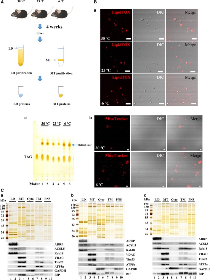
Figure 2.

Verification of purified LDs and mitochondria. LDs and mitochondria were isolated from the livers of mice after being housed at different temperatures for 4 weeks. (A) Flowchart for isolation. Briefly, LDs were isolated using sucrose density centrifugation and the resulting top layer was collected. Mitochondria were isolated using Percoll density gradient centrifugation and the resulting layer from the 50% to 25% Percoll interface was recovered. (Ba) Confocal microscopy analysis of isolated LDs by LipidTOX Red staining, DIC imaging and merged images. (Bb) Confocal microscopy analysis of isolated mitochondria by MitoTracker Red staining, DIC imaging and merged images. (Bc) Analysis of lipids extracted from isolated LDs by TLC. Scale Bar = 5 μm. (C) Silver staining and western blotting analysis of fractions from livers in mice housed at (Ca) 30 °C (Cb) 23 °C and (Cc) 6 °C. The proteins from isolated LD, MT, cytosol (Cyto), TM and PNS fractions were separated by SDS/PAGE and silver stained (upper). With equal protein loading, the indicated antibodies were tested to probe for marker proteins of different organelles/cellular fractions (lower): ADRP, ACSL5 and Rab18 (LD proteins); VDAC, Tim23 and ATP5a (MT proteins); GAPDH (cytosol protein); and BIP (ER protein).
