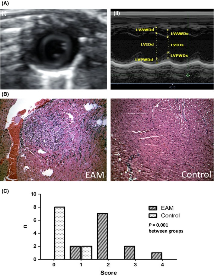
Figure 2.

A, (i) Parasternal short axis view (PSAX) of the LV at the level of the papillary muscles. (ii) M‐mode image of the PSAX with the respective measurements; LVAWDd, LV anterior wall diameter in diastole; LVAWDs, LV anterior wall diameter in systole; LVPWDd, LV posterior wall diameter in diastole; LVPWDs, LV posterior wall diameter in systole; LVIDd, LV internal diameter in diastole; LVIDs, LV internal diameter in systole. B, Animals with EAM showed significant inflammatory lesions in the histopathological examination; haematoxylin and eosin staining of mouse myocardium, magnification of 10×. C, Histogram of the myocarditis score in both groups; 0: no inflammatory infiltrates; 1: small foci of inflammatory cells between myocytes; 2: larger foci of >100 inflammatory cells; 3: < 10% of a cross‐section involved; 4: >30% of a cross‐section involved
